Environmental Medicine and Toxicology

Environmental Medicine and Toxicology

Evaluation of serum calcium, iron, and vitamin D homeostasis in relation to cataract in western Iran

Document Type : Original Article

Authors
1 Clinical Research Development Center, Imam Khomeini and Mohammad Kermanshahi and Farabi Hospitals, Kermanshah University of Medical Sciences, Kermanshah, Iran
2 Student Research Committee, Kermanshah University of Medical Sciences, Kermanshah, Iran
3 Medical Toxicology and Drug Abuse Research Center, Birjand University of Medical Sciences, Birjand, Iran
4 Substance Abuse Prevention Research Center, Research Institute for Health, Kermanshah University of Medical Sciences, Kermanshah, Iran
Abstract
Background and objectives: Cataract formation is influenced by genetic, environmental, and biochemical factors, with growing interest in the role of micronutrients like calcium, iron, and vitamin D. This study aims to examine the relationship between serum levels of these nutrients and cataract prevalence in western Iran, highlighting the potential for nutritional intervention to address this common issue.
Methods: This study involved cataract patients (n=100) in Imam Khomeini Hospital, Kermanshah, and a control group (n=100) with similar lifestyle factors. Demographic information and medical histories were obtained through a standardized checklist. Red blood cell indices, serum iron, calcium, and vitamin D levels were determined and compared in both groups. Statistical analysis was performed using R software.
Results: The study found no significant differences in vitamin D, calcium, iron, hemoglobin (Hb), and hematocrit (Hct) levels between the groups (all p > 0.05). Type 2 diabetes (OR = 3.19; 95% CI: 1.46, 6.99; p = 0.004) and hypertension (OR = 2.66; 95% CI: 1.18, 6.22; p = 0.020) were significantly associated with having cataract. Conversely, higher Hct concentrations (OR= 0.58; 95% CI: 0.45, 0.73; p < 0.001) were correlated with reduced odds of cataracts. Calcium, vitamin D, and iron levels did not show significant associations with cataracts (all p > 0.05).
Conclusion: Our study found lower hematocrit levels, type 2 diabetes mellitus, and hypertension as significant factors associated with increased odds of cataract.

Graphical Abstract

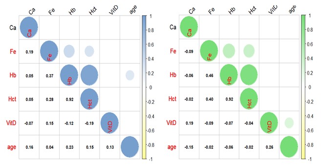
Evaluation of serum calcium, iron, and vitamin D homeostasis in relation to cataract in western Iran

Highlights

  • Type 2 diabetes and hypertension significantly increased cataract risk in Western Iranian patients.
  • Higher hematocrit levels were associated with a protective effect against cataract development.
  • Serum calcium, iron, and vitamin D levels showed no significant association with cataract.
  • Metabolic health and hematological status may be more important than micronutrient levels in cataract prevention.

Keywords
Subjects
[1] Prokofyeva E, Wegener A, Zrenner E. Cataract prevalence and prevention in Europe: a literature review. Acta ophthalmol. 2013. 91(5): 395-405. https://doi.org/10.1111/j.1755-3768.2012.02444.x
[2] Javadi MA, Zarei-Ghanavati S. Cataracts in diabetic patients: a review article. J Ophthal Vis Res. 2008. 3(1): 52-65.
[3] Abdellah MM, Mostafa EM, Salama EH, Mohamed ER. Association of Serum 25‐Hydroxyl Vitamin D Deficiency and Age‐Related Cataract: A Case‐Control Study. J Ophthalmol, 2019. 2019(1): p. 9312929. https://doi.org/10.1155/2019/9312929
[4] Atalay K, Savur FG, Kirgiz A, Kaldirim HE, Zengi O. Serum vitamin D levels in different morphologic forms of age-related cataract. Acta Endocrinol (Buchar), 2020. 16(2): 178-182. https://doi.org/10.4183/aeb.2020.178
[5] Aydin, E., Cumurcu, T., Özuĝurlu, F. et al. Levels of iron, zinc, and copper in aqueous humor, lens, and serum in nondiabetic and diabetic patients. Biol Trace Elem Res.2005. 108: 33-41. https://doi.org/10.1385/BTER:108:1-3:033
[6] Cekiç O, Bardak Y, Totan Y, Kavakli S, Akyol O, Ozdemir O, Karel F. Nickel, chromium, manganese, iron and aluminum levels in human cataractous and normal lenses. Ophthalmic Res. 1999; 31(5): 332-326. https://doi.org/10.1159/000055555
[7] Dawczynski J, Blum M, Winnefeld K, Strobel J. Increased content of zinc and iron in human cataractous lenses. Biol Trace Elem Res. 2002 Winter;90(1-3):15-23. https://doi.org/10.1385/BTER:90:1-3:15
[8] Sella R, Afshari NA. Nutritional effect on age-related cataract formation and progression. Curr Opin Ophthalmol. 2019; 30(1): 63-69. https://doi.org/10.1097/ICU.0000000000000537
[9] Jue Z, Xu Z, Yuen VL, Chan ODS, Yam JC. Association between vitamin D level and cataract: A systematic review and meta-analysis. Graefes Arch Clin Exp Ophthalmol. 2025; 263(1): 147-156. https://doi.org/10.1007/s00417-024-06592-w
[10] Allen D. Cataract. BMJ Clinical Eevidence, 2011. 2011: 0708.
[11] Theobald HE. Dietary calcium and health. Nutrit Bull. 2005. 30(3): 237-277.  https://doi.org/10.1111/j.1467-3010.2005.00514.x
[12] Pravina, P., D. Sayaji, and M. Avinash, Calcium and its role in human body. Int J Res Pharm Biomed Sci, 2013. 4(2): 659-668.
[13] Vu JT, Wang E, Wu J, Sun YJ, Velez G, Bassuk AG, Lee SH, Mahajan VB. Calpains as mechanistic drivers and therapeutic targets for ocular disease. Trends Mol Med. 2022; 28(8): 644-661. https://doi.org/10.1016/j.molmed.2022.05.007
[14] Gao J, Sun X, Martinez-Wittinghan FJ, Gong X, White TW, Mathias RT. Connections between connexins, calcium, and cataracts in the lens. J Gen Physiol. 2004; 124(4): 289-300. https://doi.org/10.1085/jgp.200409121
[15] Millonig G, Muckenthaler MU, Mueller S. Hyperferritinaemia-cataract syndrome: worldwide mutations and phenotype of an increasingly diagnosed genetic disorder. Hum Genomics. 2010; 4(4): 250-62. https://doi.org/10.1186/1479-7364-4-4-250
[16] García-Castiñeiras S. Iron, the retina and the lens: a focused review. Exp Eye Res. 2010; 90(6): 664-78. https://doi.org/10.1016/j.exer.2010.03.003
[17] Theil EC. Ferritin: at the crossroads of iron and oxygen metabolism. J Nutr. 2003; 133(5 Suppl 1): 1549S-53S. https://doi.org/10.1093/jn/133.5.1549S
[18] Micun Z, Falkowska M, Młynarczyk M, Kochanowicz J, Socha K, Konopińska J. Levels of trace elements in the lens, aqueous humour, and plasma of cataractous patients-A narrative review. Int J Environ Res Public Health. 2022 20; 19(16): 10376. https://doi.org/10.3390/ijerph191610376
[19] Brown CJ, Akaichi F. Vitamin D deficiency and posterior subcapsular cataract. Clin Ophthalmol. 2015; 9: 1093-1098. https://doi.org/10.2147/OPTH.S84790
[20] Öktem Ç, Aslan F. Vitamin D Levels in Young Adult Cataract Patients: A Case-Control Study. Ophthalmic Res. 2021; 64(1): 116-120. https://doi.org/10.1159/000509602
[21] Rim TH, Kim MH, Kim WC, Kim TI, Kim EK. Cataract subtype risk factors identified from the Korea National Health and Nutrition Examination survey 2008-2010. BMC Ophthalmol. 2014; 14: 4. https://doi.org/10.1186/1471-2415-14-4
[22] Park SJ, Lee JH, Kang SW, Hyon JY, Park KH. Cataract and cataract surgery: nationwide prevalence and clinical determinants. J Korean Med Sci. 2016; 31(6): 963-71. https://doi.org/10.3346/jkms.2016.31.6.963
[23] Foster PJ, Wong TY, Machin D, Johnson GJ, Seah SK. Risk factors for nuclear, cortical and posterior subcapsular cataracts in the Chinese population of Singapore: the Tanjong Pagar Survey. Br J Ophthalmol. 2003; 87(9): 1112-1120. https://doi.org/10.1136/bjo.87.9.1112
[24] Liu H, Shen X, Yu T, Wang Y, Cai S, Jiang X, Cai X. A putative causality of vitamin D in common diseases: A mendelian randomization study. Front Nutr. 2022; 9: 938356. https://doi.org/10.3389/fnut.2022.938356
[25] Jee D, Kim EC. Association between serum 25-hydroxyvitamin D levels and age-related cataracts. J Cataract Refract Surg. 2015; 41(8): 1705-1715. https://doi.org/10.1016/j.jcrs.2014.12.052
[26] Park S, Choi NK. Serum 25-hydroxyvitamin D and Age-Related Cataract. Ophthalmic Epidemiol. 2017; 24(5): 281-286. https://doi.org/10.1080/09286586.2017.1281427
[27] Aidenloo NS, Motarjemizadeh Q, Nahriq SM. Serum concentrations of 25-hydroxyvitamin D and different subtypes of age-related cataract; a case-control study. Immunopathologia Persa. 2021 Feb 22;8(1):e16-.
[28] Rao P, Millen AE, Meyers KJ, Liu Z, Voland R, Sondel S, Tinker L, Wallace RB, Blodi BA, Binkley N, Sarto G, Robinson J, LeBlanc E, Mares JA. The Relationship Between Serum 25-Hydroxyvitamin D Levels and Nuclear Cataract in the Carotenoid Age-Related Eye Study (CAREDS), an Ancillary Study of the Women's Health Initiative. Invest Ophthalmol Vis Sci. 2015; 56(8): 4221-4230. https://doi.org/10.1167/iovs.15-16835
[29] Appleby PN, Allen NE, Key TJ. Diet, vegetarianism, and cataract risk. Am J Clin Nutr. 2011; 93(5): 1128-35. https://doi.org/10.3945/ajcn.110.004028
[30] Tavani A, Negri E, La Vecchia C. Food and nutrient intake and risk of cataract. Ann Epidemiol. 1996 Jan;6(1):41-6. https://doi.org/10.1016/1047-2797(95)00099-2
[31] Rahman ST, Waterhouse M, Romero BD, Baxter C, English D, Mackey DA, et al. Vitamin D Supplementation and the Incidence of Cataract Surgery in Older Australian Adults. Ophthalmology. 2023; 130(3): 313-323. https://doi.org/10.1016/j.ophtha.2022.09.015
[32] Alvarez JA, Chowdhury R, Jones DP, Martin GS, Brigham KL, Binongo JN, et al. Vitamin D status is independently associated with plasma glutathione and cysteine thiol/disulphide redox status in adults. Clin Endocrinol (Oxf). 2014; 81(3): 458-466. https://doi.org/10.1111/cen.12449
[33] Uberti F, Lattuada D, Morsanuto V, Nava U, Bolis G, Vacca G, Squarzanti DF, Cisari C, Molinari C. Vitamin D protects human endothelial cells from oxidative stress through the autophagic and survival pathways. J Clin Endocrinol Metab. 2014; 99(4): 1367-74. https://doi.org/10.1210/jc.2013-2103
[34] Jee D, Kang S, Yuan C, Cho E, Arroyo JG; Epidemiologic Survey Committee of the Korean Ophthalmologic Society. Serum 25-Hydroxyvitamin D Levels and Dry Eye Syndrome: Differential Effects of Vitamin D on Ocular Diseases. PLoS One. 2016 19; 11(2): e0149294. https://doi.org/10.1371/journal.pone.0149294
[35] Chattopadhyay N, Ye C, Singh DP, Kifor O, Vassilev PM, Shinohara T, Chylack LT Jr, Brown EM. Expression of extracellular calcium-sensing receptor by human lens epithelial cells. Biochem Biophys Res Commun. 1997; 233(3): 801-805. https://doi.org/10.1006/bbrc.1997.6553
[36] Velpandian T, Nirmal J, Gupta P, Vijayakumar AR, Ghose S. Evaluation of calcium dobesilate for its anti-cataract potential in experimental animal models. Methods Find Exp Clin Pharmacol. 2010; 32(3): 171-179. https://doi.org/10.1358/mf.2010.32.3.1423888
[37] Gupta PD, Johar K, Vasavada A. Causative and preventive action of calcium in cataracto-genesis. Acta Pharmacol Sin. 2004 Oct;25(10):1250-6.
[38] Duncan G, Wormstone IM. Calcium cell signalling and cataract: role of the endoplasmic reticulum. Eye (Lond). 1999; 13( Pt 3b): 480-483. https://doi.org/10.1038/eye.1999.125
[39] Raman R, Pal SS, Adams JS, Rani PK, Vaitheeswaran K, Sharma T. Prevalence and risk factors for cataract in diabetes: Sankara Nethralaya Diabetic Retinopathy Epidemiology and Molecular Genetics Study, report no. 17. Invest Ophthalmol Vis Sci. 2010; 51(12):6253-62561. https://doi.org/10.1167/iovs.10-5414
[40] Tarwadi K, Agte V. Linkages of antioxidant, micronutrient, and socioeconomic status with the degree of oxidative stress and lens opacity in indian cataract patients. Nutrition. 2004; 20(3): 261-267. https://doi.org/10.1016/j.nut.2003.11.020
[41] Garland D. Role of site-specific, metal-catalyzed oxidation in lens aging and cataract: a hypothesis. Exp Eye Res. 1990;50(6):677-82. https://doi.org/10.1016/0014-4835(90)90113-9
[42] Nam SW, Lim DH, Cho KY, Kim HS, Kim K, Chung TY. Risk factors of presenile nuclear cataract in health screening study. BMC Ophthalmol. 2018; 18(1): 263. https://doi.org/10.1186/s12886-018-0928-6
Volume 1, Issue 1
Summer 2026
Pages 31-40

  • Receive Date 09 September 2026
  • Revise Date 18 September 2026
  • Accept Date 24 September 2026
  • First Publish Date 24 September 2026
  • Publish Date 27 September 2026